 »
Danh Mục Bài Viết
»
Kỹ thuật phẫu thuật
»
Danh Mục Bài Viết
»
Kỹ thuật phẫu thuật
Laparoscopic-Assisted Right Colectomy (kỹ thuật cắt đại tràng phải nội soi hỗ trợ)
Thứ bảy - 25/06/2011 15:15
Laparoscopic Right Colectomy
Step 1.
After general anesthesia, place the patient in “laparoscopic” lithotomy position and place a nasogastric tube and Foley catheter.
Step 2.
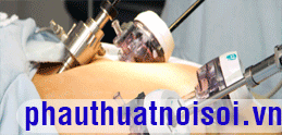
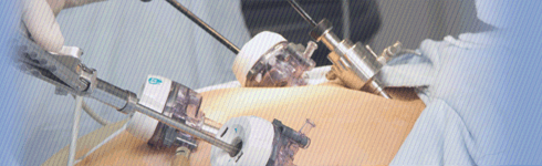
Make a periumbilical incision for GelPort placement (Fig. 1 ). This allows initial open dissection of the transverse colon and entry to the lesser sac. Depending on the patient’s body habitusa great deal of dissection can be done through this open incision, in cluding transection of the mesocolon, lysis of adhesions, and separation of any inflammatory adhesions. Place 5-mm ports in the suprapubic and left upper quadrant positions. Optional 5-mm ports can be placed midline and subxiphoid. Place a 10-mm port in the right lower quadrant.

Figure 1
Step 3.
The surgeon stands on the patient’s left along with the assistant.
Step 4.
With the patient in Trendelenburg position and left side down the surgeon grasps the cecum and right colon retracting it toward the patient’s left. The Harmonic Scalpel is used to incise along the white line of Toldt caudally and cephalad. Perform this dissection up to and around the hepatic flexure. For easy dissection, visualization, and broad planes of dissection it is essential that the surgeon’s index finger is strategically placed underneath the lateral attachments of the colon and the hepatic colic ligament. The assistant retracts the omentum cephalad during the hepatic flexure mobilization. The dissection proceeds from lateral to medial. During this phase all small bowel is manipulated to the left upper quadrant. Carefullyidentify the ureter and gonadal vessels. This portion of the dissection is complete when the duodenum is visualized. Continue the dissection toward the patient’s lesser sac, dissecting the avascular plane between the omentum and transverse colon.
Step 5.
Ligate the vessels with the endoscopic GIA, using either 30 mm or 45 mm vascular staples. Quite often the stapler is not necessary. With good mobilization the mesocolon can be divided through the periumbilical incision.
Step 6.
Bring out the transverse colon and ileum through the periumbilical incision. During this phase, the GelPort acts as a wound retractor and protector. Perform a stapled anastomosis using a 75-mm GIA and 60-mm TA. The mesentery is reapproximated, whereas in the left colectomy it is not.
Step 7.
Close the midline wound with 1 PDS. Close the skin with 4-0 Vicryl.
Tác giả bài viết: Dr. Ai
 Xem phản hồi
 Gửi phản hồi
Những tin mới hơn
Những tin cũ hơn
ĐĂNG NHẬP
CÁC BỆNH THƯỜNG GẶP
- BỆNH THOÁT VỊ BẸN
Tư Vấn Trực Tuyến
Thống kê truy cập
![]() Đang truy cập :
42
Đang truy cập :
42
•Máy chủ tìm kiếm : 16
•Khách viếng thăm : 26
![]() Hôm nay :
3318
Hôm nay :
3318
![]() Tháng hiện tại
: 107636
Tháng hiện tại
: 107636
![]() Tổng lượt truy cập : 57440428
Tổng lượt truy cập : 57440428